Minds On
Organ systems
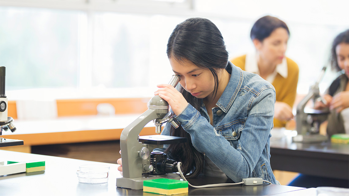
In this learning activity, you will be exploring different organisms, including human organ systems, and making connections between the human body, cell research, and advancements.
Before you begin, complete the following matching activity to review the human organ systems and their functions.
For each system, select the corresponding function.
Brainstorm
Brainstorm
What are some of the individual organs that you might find in each system?
Record your ideas in a notebook or another method of your choice.
If possible, share your ideas with a partner.
Action
Cells
The body is a living system that is made up of many different parts that work together to form a function. In previous grades, you likely learned that human organ systems play an important role in keeping people alive and maintaining their health.
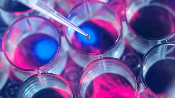
Scientists have been studying living systems for many years, including exploring cells. Once scientists realized that all living things are made up of cells, they developed the cell theory.
Cell theory
Cell theory explains the basic characteristics of all living things.
- All living things are composed of one or more cells.
- The cell is the basic unit of life.
- All cells come from pre-existing cells.
Studying cells helps us to understand how all living things work.
Unicellular or multicellular
An organism is either unicellular or multicellular.
Press the following tabs to access these key terms.
A unicellular organism (for example, yeast, algae, and some fungi) only relies on one cell to carry out all of the necessary functions of an organism.
A multicellular organism is much more complex and requires a variety of different cells to function. For example, the human body requires blood cells, nerve cells, skin cells, and muscle cells to survive.
Multicellular organisms
At each level of organization – cells, tissues, organs, and organ systems – structure is closely related to function.
The first level or organization in multicellular organisms is cells, then tissues, organs, and lastly, organ systems.

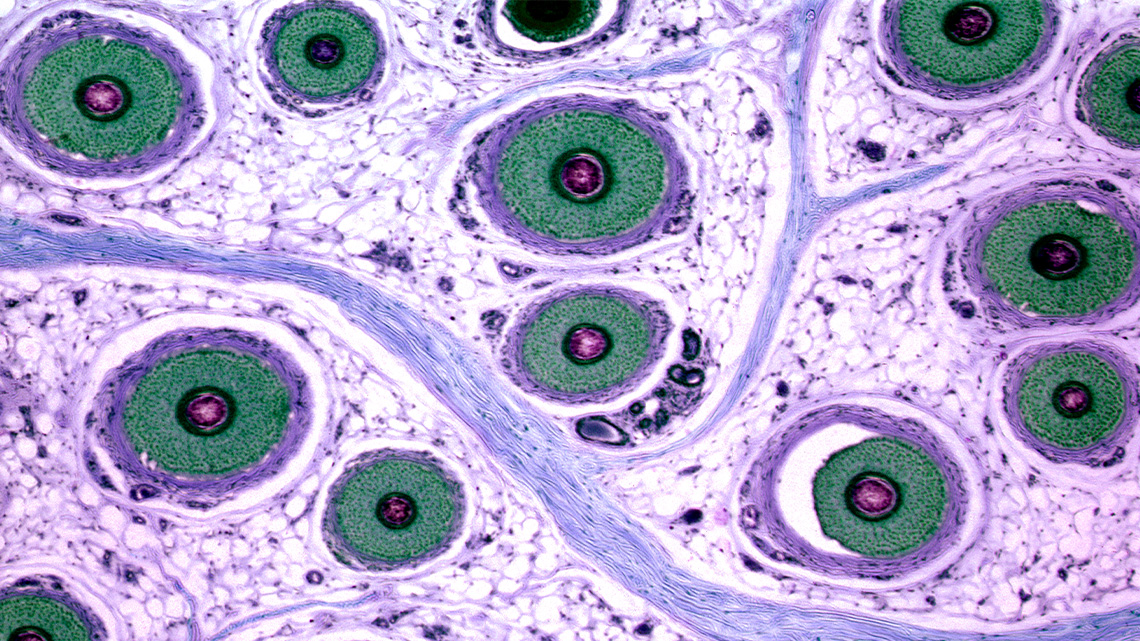
Tissue
The second level of organization in a multicellular organism is tissue.
Tissue is a group of cells that have similar shape and function. Organs are composed of groups of tissues, and organ systems are composed of groups of organs.
Press the following tabs to access the four types of tissues.

Epithelial tissue
Epithelial tissue consists of tightly packed sheets of cells that cover surfaces, like the outside of the body.

Connective tissue
Connective tissue is responsible for supporting and connecting other tissues. These cells are suspended in a jelly-like substance.

Muscle tissue
Muscle tissue is essential for movement, pumping blood, and digestive tract contraction. Certain proteins allow the cells to contract.

Nervous tissue
Nervous tissue consists of nerve cells and glia. It senses internal/external cues to process and transmit information.
Did You Know?
Did you know?
When the tissues are grouped together, they perform a complex task.
As a group, these tissues are called an organ.
An organ has muscle tissue, connective tissue, and nerve tissue all working together. As a group, these tissues pump blood.
Examples of organs include the heart, skin, lungs, and the brain.
When organs work together, they form a system in the body, like the respiratory system, muscular system, and many more!
Learning check!
For each tissue type, select the corresponding description.
Unicellular organisms
Unicellular organisms are made up of one single cell. Their function is carried out by the one cell, which, in most cases, requires a microscope to view.
Bacteria are unicellular organisms that have cell walls, but lack organelles and a nucleus. Some bacteria can cause disease.


A carousel of two images. Image 1: This image includes five unicellular organisms. They are all different in appearance. One organism is oval shaped with a dark brown circle inside and small circles throughout, close together but not touching. The second organism is a long oval shape with one pointy end. It has a thread at one end, partly inside and partly outside of the cell. There is one red circle, eight thin green sticks, seven tiny circles and one yellow circle with short lines surrounding it. The third organism is a green circle with a red circle, two yellow thin oval objects and a green oval with five small ovals inside. The fourth organism is represented as a starfish shape with circles of various sizes inside. There is a red circle in the centre and one thin purple object inside. The fifth organism is a long oval shape with one pointy end. It has small lines surrounding the organism on the outside. Inside are five small circles, one large blue oval shaped object a red object attached to the inside of the organism wall, and two small yellow circles with short lines surrounding each. Image 2: This bacterial cell is represented as a green oval shaped object with a long thread, labelled as a flagella on one end. Surrounding the outside of the cell are short threads surrounding it, labelled pilus. On the inside is the space before the cell wall labelled the capsule. Then there is a blue line representing the cell wall. Inside the cell wall is a large orange object which is labelled the nucleoid. There are small blue circles labelled ribosomes, a larger blue circle labelled a food granule, a collection of thin oval shapes labelled mesosome and a ring that is labelled plasmid.
According to the National Academy of Sciences, one single human body is said to have around one hundred trillion individual bacteria cells. Although many types of bacteria can cause human and animal diseases, some bacteria can be very important, and some have been known to help in agriculture and the various food industries.
Investigate
Investigate
Let’s explore and research the benefits and risks of bacteria.
Before beginning your research, explore this video to learn about the steps of the Scientific Research Process.
As you are researching, record your research findings and ideas in the following organizer or another method of your choice.
Complete the Benefits and Risks of Bacteria Activity in your notebook or using the following fillable and printable document. If you would like, you can use speech-to-text or audio recording tools to record your thoughts.
|
Bacteria benefits |
Bacteria risks |
|---|---|
Press the ‘Activity’ button to access the Benefits and Risks of Bacteria Activity.
When you’re ready, compare your findings to the answer key. Access the following Answers to Benefits and Risks of Bacteria to review your findings.
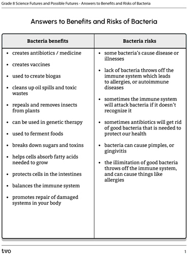
Press the Activity button to access the Answers to Benefits and Risks of Bacteria.
Activity (Open PDF in a new tab)Future impacts
This learning activity connects new and existing approaches for young scientists to create positive changes in their communities.

Cells are very important because they are the basic building blocks of all living things. Studying cells helps us understand how organisms function, and how organs work together to carry out basic functions.
Diseases

Scientists use their knowledge and research about cells to treat illnesses and diagnose diseases. Scientific research has led to discoveries about different activities that harm and benefit cells.
For example, scientists have discovered that ultraviolet light and other radiations can damage DNA, which leads to cancer. Cancer happens when abnormal and harmful cells grow and divide and do not get destroyed. Research has also led to discoveries on how we can keep our cells healthy with the proper vitamins, minerals, and fatty acids needed to survive and thrive.
Innovations
There have been several innovative technologies and contributions to cell discoveries over the years.
Press the following tabs to explore some of the technologies available now and in the years to come.
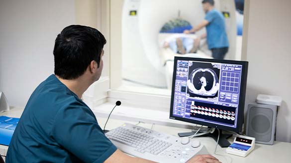
A Positron Emission Tomography (PET) scan is an innovative scanning technology used to picture what is happening inside the body at a cellular level. This type of scan offers a unique insight into the human body and allows doctors to personalize patient care.
These scans help doctors:
- determine the exact location and severity of a disease
- select the most effective therapy and treatment for the disease
- determine a patient's response to specific treatments
- assess disease progression
When a disease occurs, such as cancer or dementia, abnormal cellular activity affects tissues and structures, which can be detected using a PET scan. PET scans are very useful because they can detect cellular changes early on.
So, how does a PET scan work? A special dye that uses a radioactive tracer (swallowed, inhaled, or injected) will collect in areas where there is higher chemical activity, which will show up on the PET scan imaging. It then creates a 3D image that can be studied for diseases, and to determine treatment plans.
Most of the cells in the human body are specialized, meaning they perform a specific function in a certain kind of tissue.
Stem cells are considered unspecialized cells of the human body, which means they can renew themselves and take on many different cell roles. The ways in which stem cells can be developed into specialized cells are still being investigated by scientists, and have yet to be fully understood.
Scientists are working to develop strategies that use stem cells to replace defective or damaged cells in the body. This makes stem cell research one of the most important components of modern and future medicine. These cells could be used to treat a variety of diseases and injuries, such as Alzheimer’s disease, Parkinson’s disease, heart disease, and diabetes.
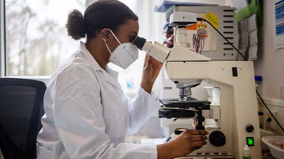
Around the world, teams of scientists, engineers, and technicians are working to take microscopy to the next level.
Microscopy is the technical field of using microscopes to view objects and areas of objects that cannot be observed using the eye alone.
Electron tomography scans untreated samples of cells and tissues with beams of electrons to create 3D images using a computer. So far, one drawback is that only deceased specimens can be viewed, however, it is believed that scientists will discover a way to use live specimens in the future.
Engineers are continually developing better tools for investigating cells. As new microscopes are invented, valuable research will continue to expand.
Check your understanding of specialized cells, scanning cells, and microscopy.
Select the correct answer, then press ‘Check Answer’ to see how you did.
Consolidation
Review your learning
The levels of organization and structure are closely related to function. Place the levels in each answer box in the correct order.
Student Success
Think
Reflect on the following questions:
- Why is it important to study cells for the future? How is the study of cells impacting our future and changing our lives?
- What is the difference between a unicellular and multicellular organism?
Create a sorting activity about unicellular and multicellular organisms. Be sure to include an answer key. If possible, you may share your activity with a partner or teacher.
Record your ideas and thoughts in a notebook or another method of your choice.

Reflection
As you read the following descriptions, select the one that best describes your current understanding of the learning in this activity. Press the corresponding button once you have made your choice.
I feel…
Now, expand on your ideas by recording your thoughts using a voice recorder, speech-to-text, or writing tool.
When you review your notes on this learning activity later, reflect on whether you would select a different description based on your further review of the material in this learning activity.
Press ‘Discover More’ to extend your skills.
Discover MoreCareer connections
So, who does the work of creating innovations in the study of cells, anyway?
-
Choose one of the following careers to investigate further:
- bioengineering researcher
- biomedical research assistant
- cancer researcher
- cellular engineering researcher
-
Respond to the following questions:
- What kind of work do they do that involves cells?
- What education is required for this career?
- How are they helping people?
Record your ideas in a notebook or another method of your choice.