Minds On
What is a cell?

Do you know what a cell is? Do you know what cells do or how many different types there are? Why are they so important to living things?
Record your thoughts to these questions using the following Mind Map organizer or using another method of your choice.
You may complete the Mind Map in your notebook or using the following fillable and printable document. If you would like, you can use speech-to-text or audio recording tools to record your thoughts.
Agree or disagree?
Next, explore the following statements and rank them using the following 1-4 rating system. Record your thoughts using a method of your choice.
- Some cells are constantly being replaced.
- Some cells last an entire lifetime.
- The new cells need energy to grow.
Rating system:
1 = strongly disagree
2 = disagree
3 = agree
4 = strongly agree
Action
Cells, cells, cells!
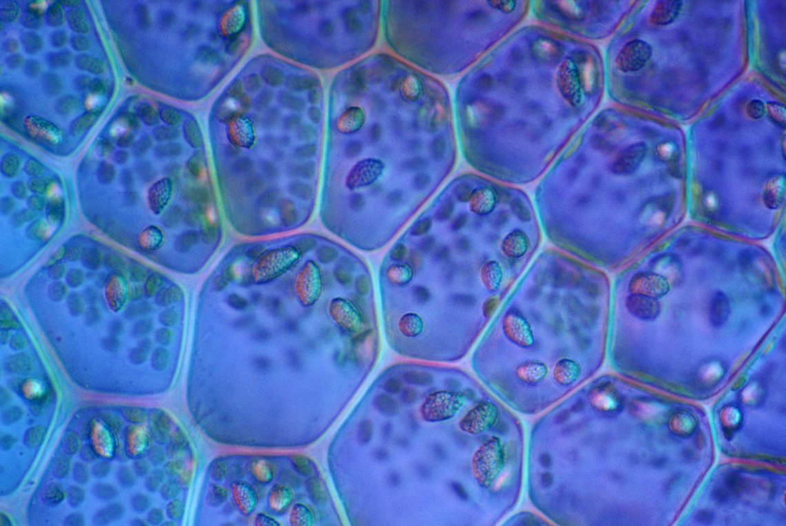
Animal and plant cells
Every living thing is made up of cells. To be considered a living thing, an organism must possess the following things:
- Nutrition–they must obtain energy from something
- Respiration–they must release some form of energy
- Excretion–they must expel waste
- Sensitivity–they must detect change in their environment
- Growth–they must grow
- Reproduction–they must reproduce and pass their traits on to their offspring
- Movement–they must move in some capacity
Animal Cells are round, smaller in size with small vacuoles and do not have cell walls.

An animal cell with labelled parts. The parts of the cell include: centrosome, cytoplasm, mitochondria, rough endoplasmic reticulum, smooth endoplasmic reticulum, nucleus, Golgi apparatus, ribosomes, vacuole, lysosome, and the cell membrane.
Plant Cells are square, larger in size with large vacuoles and a cell wall. They have chloroplasts for photosynthesis.

A plant cell with labelled parts. The parts of the cell include: Golgi apparatus, cytosol, endoplasmic reticulum, nucleus, vacuole, chloroplast, mitochondria, the cell membrane, and the cell wall.
Explore the following set of flashcards to learn about the structures and functions of organelles found only in animal cells, only in plant cells, or found in both!
Explore the next set of flashcards to learn more structures and functions of organelles that are found in both animal and plant cells.
Student Success
Create your own resource
In your notebook or digitally, create your own flashcards, a visual glossary, or study notes/diagrams for each of the cell organelles presented. Choose the format that works best for you to use as a resource to review and study the structures and functions of each of the organelles.
Cell theory
There are 6 principles of cell theory:
- All living things are composed of cells.
- The cell is the basic unit of life.
- All cells come from pre-existing cells.
- Energy flow occurs within cells.
- DNA is contained in cells.
- All cells have the same basic chemical composition.
In this section of the learning activity, you are going to explore one of the components of cell theory: cellular reproduction.
Cellular reproduction
Why do cells constantly need to reproduce? Firstly, new cells are needed in order for organisms to grow – for an organism to get larger, new cells are needed. Secondly, cells reproduce to replace old or damaged cells. A cell can become damaged from heat, physical contact, and lack of nutrients or oxygen. If a cell is damaged, the organism will break it down, get rid of it, and replace it. A real-life example of this is when someone cuts themselves, the tissue needs to heal and overtime cells in the tissue will be replaced by new cells as they heal.
So, how do cells reproduce? Explore Mitosis to learn more!
Mitosis
The process that the majority of cells undertake to reproduce is called Mitosis.
Press the following tabs to learn about each step of Mitosis.
DNA starts to form two sets of identical chromosomes on the inside of the nucleus.

This image of prophase shows a circular shape with a circle inside that includes 6 X shaped objects, 3 are red in colour and 3 are blue. In this circular shape are also 2 yellow objects with yellow lines surrounding them.
The nuclear membrane starts to dissolve (Notice the circle inside the cell has disappeared.)

This image of prometaphase shows a circular shape with 6 X shaped objects inside in the centre of the shape. 3 are red and 3 are blue. In this circular shape are also 2 yellow objects with yellow lines surrounding the object throughout the circular shape.
Parts, known as “spindles” attach to the chromosomes (notice how the x’s are attaching together.)

This image of metaphase shows a circular shape with 6 X shaped objects lined up one on top of the other through the centre of the circle. Two blue, two red, then one blue, then one red. On either side of this line is a yellow object with yellow lines surrounding the object throughout the circular shape.
The chromosome pairs are pulled apart (notice the x’s are pulled in half and in different directions.)

This image of anaphase shows two circular shapes attached together. Each shape has 6 V shaped objects inside, one under the other in a line along one side of the circle. There are yellow lines that reach from each V shape to a singular yellow object on the other side of each circle. The V shaped objects are the same in each circle in terms of colour pattern. Two blue, then two red, then one blue, then one red.
New nuclei start to form, and all other parts of the cell are divided in two halves.

This image of telophase and cytokinesis shows two circular shapes attached together. Each shape has another dark purple circle within it, about half the size of the shape. In these purple circles are 6 thin objects, 3 red and three blue. There is also a small blue circle with these shapes. Outside of the dark purple circle in each circular shape is a small yellow object with yellow lines surrounding it.
The cell membrane pinches closed in the middle and two separate cells are formed.

In this Telophase and Cytokinesis diagram are two circular shapes, one above the other. Each shape has another dark purple circle within it, about half the size of the shape. In these purple circles are 6 thin objects, 3 red and three blue. There is also a small blue circle with these shapes. Outside of the dark purple circle in each circular shape is a small yellow object with yellow lines surrounding it.
For each sentence, select the missing word from the drop-down menu.
Exploring diffusion and osmosis
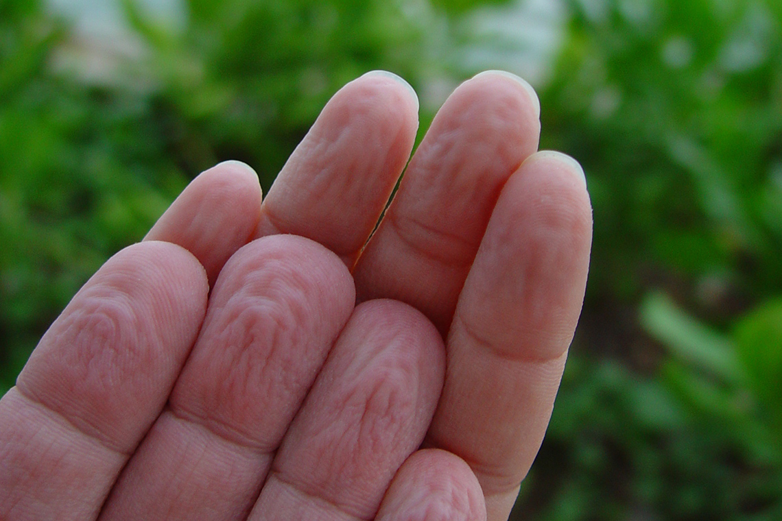
Why do fingers wrinkle when they have been in water for too long?
Brainstorm your ideas using a method of your choice, such as in print, digitally or with an audio recording. If possible, share your ideas with someone else.
Press “Answer” to access an explanation of why fingers wrinkle when placed in water for a long time.
If a person stays in water for too long, water will begin to flow into the upper skin cells causing visible wrinkles – this is an example of a process called osmosis. Shrinking and expanding effects take place at the same time in these skin cells, which causes wrinkles.
Movement through membranes
As you learned earlier, the cell membrane acts like a gatekeeper and controls what can enter and exit the cell. Examine the following definitions to begin to explore how different particles move through membranes in cells.
- Semi Permeable Membrane is a membrane that allows certain molecules or ions to pass through it by diffusion or osmosis
- Diffusion is the movement of particles at random from an area of higher concentration to an area of lower concentration
- Osmosis is the movement of water molecules through a selectively permeable membrane from an area of high concentration to an area of lower concentration
Explore the following animation to examine the difference between diffusion and osmosis. Can you note the difference?

To further explore the differences between diffusion and osmosis, you have two options to choose from, or you can complete both!
- Option one: complete a hands-on experiment to explore diffusion and osmosis
- Option two: examine and record observations of a video demonstration from Science North of experiments that demonstrate osmosis and diffusion.
Before you choose which option you will complete, let’s first explore a video that explains the Scientific Experimentation Process. This will help guide you as you work your way through your choice of activity.
Check out this video to learn about the steps of the Scientific Experimentation Process.
Option one: hands-on experiment
It’s time to experiment!
Before you begin, let’s perform a safety check.
Safety
Explore the following list to perform your safety check.
Hands-on Science
Hands-on science
For this experiment, we will be exploring the differences between diffusion and osmosis.
Complete the Lab Sheet: Osmosis and Diffusion in your notebook or using the following fillable and printable document. If you would like, you can use speech-to-text or audio recording tools to record your thoughts.
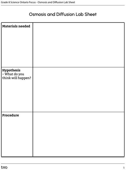
Press the Activity button to access the Lab Sheet: Osmosis and Diffusion.
Activity (Open PDF in a new tab)Press the following tabs to access the materials and steps for the Osmosis and Diffusion experiment.
If you do not have access to materials, access the “Video demonstration” tab to explore the experiment in action. You can use the video to make your observations and draw your conclusions.
Part A: osmosis
- 3 clear containers (jars or cups)
- 3 gummy animal candies (each a different colour)
- Tap or bottled water
- Plate (to set wet gummy candies on, preferably white)
- Spoon or fork to take gummy candies out
- Clock or timer
- Ruler (to measure gummy candies)
- Pencil or pen to record your observations
Part B: diffusion
- Plate (preferably white and not paper)
- Bag of hard-shelled candies
- Very warm water
- Pencil or pen to record your observations
Part A: osmosis
- Measure the length, width and height of the gummy candies one at a time and record them in the observations chart provided.
- Fill the three cups of water up evenly.
- Add one gummy candy to each cup of water.
- Record your observations after 2, 4 and 6 hours on the observations chart. Do not remove the gummy candies. (NOTE: 6 hour time can be adjusted to longer if experiment runs overnight. Just adjust chart to new length of time).
- After 6 hours, remove each gummy candy individually and place them on the plate.
- Measure the length, width and height of the three gummy candies again and record the data in the observations chart. What did you notice?
Part B: diffusion
- Place the hard-shelled candies around the edge of the plate, trying to alternate the different colours.
- Slowly pour the very warm water in the center of the plate until it just covers the bottom of all the candies. Record what happens in the observations chart.
Check out this video to explore a demonstration of the Osmosis and Diffusion experiment. Please note that materials and procedure in the video may vary slightly from what is listed in the previous tabs.

Science is about reflecting and reimagining. Was your experiment successful?
Is there anything that you would change about your experiment design to improve it or the outcome?
Even if your experiment was not successful, what did you learn or confirm about the topic you were investigating?
Press ‘Conclusions’ to access some conclusions from the experiment.
Through osmosis, the tap water that was originally outside the gummy bear travelled through the semi permeable membrane of the gummy bear and caused it to grow.
The molecular structure of the dyes used on the skittles makes it easy for them to dissolve in warm water and then diffuse.
Both diffusion and osmosis aim to equalize forces inside cells and organisms, spreading water, nutrients and necessary chemicals from areas that contain a high concentration to areas that contain a low concentration.
Option two: observing demonstrations
As you explore the demonstrations of diffusion and osmosis, complete the provided Notice, Think, Wonder Chart, or use another method of your choice to record your observations.
Complete the Notice, Think, Wonder Chart in your notebook or using the following fillable and printable document. If you would like, you can use speech-to-text or audio recording tools to record your thoughts.
|
I notice… (only record things you can actually notice yourself) |
|
|
I think… (record what you think from what you’ve noticed and what you might already know) |
|
|
I wonder… (record all of the questions you have) |
Press the ‘Activity’ button to access Notice, Think, Wonder Chart.
Explore the following video from Science North to record your observations of the differences between diffusion and osmosis.
Forming conclusions
After completing the hands-on experiment, observing the video demonstrations, or both, what conclusions can you make about the differences between diffusion and osmosis in cells? Are there any similarities? Can you explain how each works within cells?
Record your thinking using a method of your choice such as in print, digitally or using an audio recording.
Press “Hint” to access some information to help you form your conclusions.
Consolidation
Review your learning

Complete the following tasks.
Task 1: matching!
For each term select the corresponding definition.
Task 2: diffusion and osmosis
Create a visual diagram or detailed description that explains the difference between diffusion and osmosis. Your diagram or description should include how diffusion and osmosis works from a high to low concentration and have semi-permeable membrane labelled.
Press ‘Sample Answer’ to access a sample visual diagram.

For diffusion, particles in a container are at first tightly packed, and then spread outward in all directions. The solute moves from a high to a low concentration. For osmosis, water particles in a mixture move through a semi-permeable membrane while solute particles are left behind. The solvent (water) moves from a high to a low concentration.
Task 3: compare and contrast
Record the similarities and differences between plant and animal cells. You should use some of the key words from the previous definitions activity to help you.
Complete the Venn Diagram in your notebook or using the following fillable and printable document. If you would like, you can use speech-to-text or audio recording tools to record your thoughts.
Press ‘Sample Answer’ to access a possible answer to the Venn Diagram.

A Venn diagram for animal cells and plant cells. The animal cell section has the following: Smaller, Smaller vacuole, Cell wall is absent, round shape. The plant cell section has the following: Larger, larger vacuole, has rigid cell wall, rectangular shape, have chloroplasts for photsynthesis. In the both section is the following: Vacuole, Golgi apparatus, Mitochondria, Smooth endoplasmic reticulum, Rough endoplasmic reticulum, Cytoplasm, Cell membrane, Ribosomes, and Nucleus.
Reflection
As you read the following descriptions, select the one that best describes your current understanding of the learning in this activity. Press the corresponding button once you have made your choice.
I feel…
Now, expand on your ideas by recording your thoughts using a voice recorder, speech-to-text, or writing tool.
When you review your notes on this learning activity later, reflect on whether you would select a different description based on your further review of the material in this learning activity.