Minds On
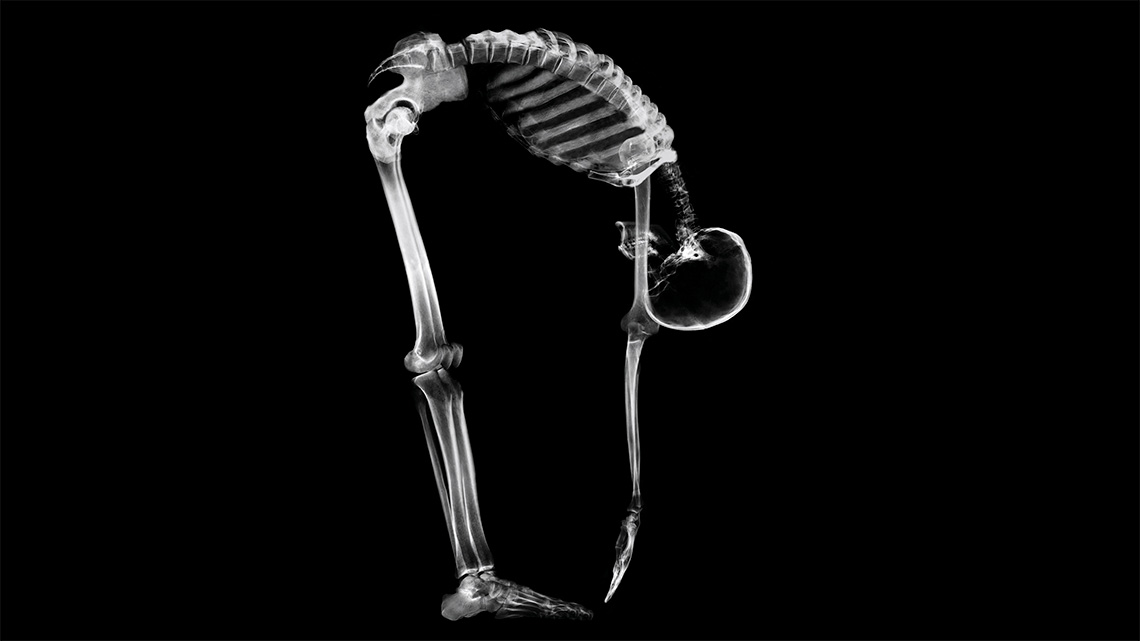
The human body
The human body is made up of many cells and systems that work together. Can you think of any systems that help the human body function? Explore the images to help you.
Record your list in a method of your choice.

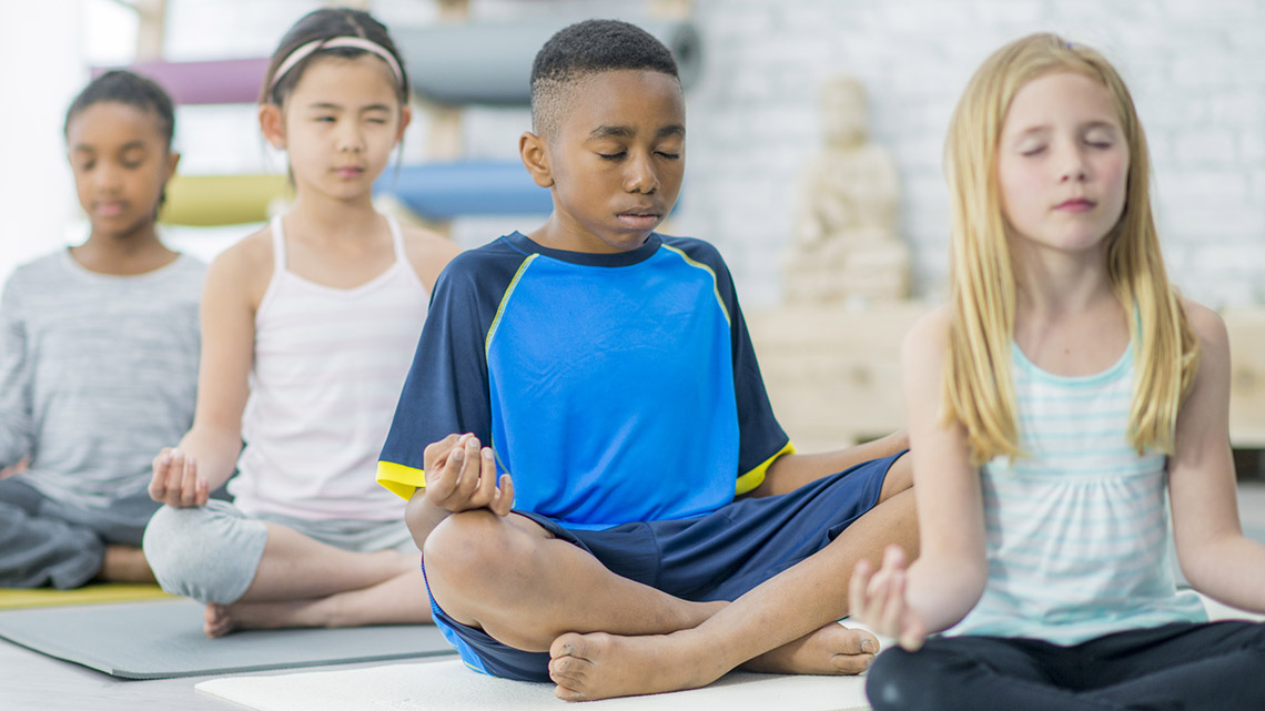
Children sitting on mats with their legs crossed, their hands on their knees, their eyes closed and breathing in and out.
Action
Different body systems
Human bodies are made of cells which are specialized in their function to support the work of different organs. Examples of organs include the heart, lungs, stomach, liver, pancreas and kidneys.
Some organs are organized together into systems.
As you move throughout this learning activity, you may use the following organizer to record a definition of each system in your own words as well as describe the parts of the body involved and the purpose of the system.
Complete the Body Systems Chart in your notebook or using the following fillable and printable document. If you would like, you can use speech-to-text or audio recording tools to record your thoughts.
|
Body system |
Parts of the body involved |
Purpose of the body system |
|---|---|---|
Press the ‘Activity’ button to access Body Systems Chart.
Explore the following carousel of images that describe of some of the body systems.

Cardiovascular System
The cardiovascular system is made up of arteries, veins, blood and the heart. It’s function is to transport blood carrying nutrients and gases around the body.

Nervous System
The nervous system is given information by the senses of sight, hearing, taste, smell and touch and that information is sent to the brain to then decide what to do next, and signal the system of the body that should react.

Respiratory System
The respiratory system exchanges fresh oxygen from inhaling for the body’s waste product of carbon dioxide by exhaling. This system works with the cardiovascular system for delivery of oxygen and carbon dioxide.

Digestive System
The digestive system is a series of tubes that use muscular contractions to move food from one end to the other. The function is to absorb nutrients from this system into the bloodstream, and then the waste products travel out of the body.

Musculoskeletal system
The musculoskeletal system pairs a moveable internal frame that provides structure for all other systems and muscles, tendons and ligaments that support movement.
Image 1: This image of the cardiovascular system highlights the veins, capillaries and arteries that travel throughout the body from head to toe, including the arms and legs. These are represented as long red and blue tubes. The heart is also shown in this image and the veins and arteries that extend from the heart.
Image 2: This image of the nervous system highlights the brain, spinal cord travelling down the back, nerves which are represented as threads travelling throughout the body from head to toe, including arms and hands. The brain is represented as a large oval shaped object at the top half of the head.
Image 3: This image of the respiratory system highlights the nose and mouth area of the head, the nasal cavity and trachea which is a tube connecting the nose and mouth highlights to the two lungs which are oval shaped objects in the chest area of the body.
Image 4: This image of the digestive system highlights the mouth area as a circle, then a tubular esophagus running down to the stomach, and tubular shaped small intestines, large intestines.
Image 5: This image of the musculoskeletal system shows a person with half bones, half muscles. The muscle side highlights the muscles as long, thin and thick lines throughout the entire body, from head to toes and hands. The skeletal side highlights the skeleton inside the body. It includes the skull the spinal cord extending from the skull to the pelvis which is two bones at the bottom of the torso. There are bones extending from the chest to the shoulders, then down the arms into the hands and fingers. There are also bones extending from the pelvis down the leg into the feet and toes.
Let’s look closer at the digestive system.
The digestive system consists of specialized cells to help absorb nutrients from food. The organs of the digestive system include the mouth, esophagus, stomach, intestines and anus. This system is essentially one long tube designed to break food into smaller and smaller pieces so that a body can absorb nutrients.
The digestive system works closely with the cardiovascular system whose job it is to deliver the nutrients through your blood stream. The organs involve in the cardiovascular system are the heart, arteries that take blood away from the heart, veins that return blood back to the heart, and tiny capillaries where oxygen is delivered from ‘fresh’ blood and the waste product of carbon dioxide is picked up to be delivered back to the heart.
Learning check!
For each description select the corresponding system.
Task 1: Investigating with pulse

The heart is a very strong muscle that contracts automatically. Unlike if you want to move your arm, you do not need to think about making your heartbeat. The system that involves your heart is the cardiovascular system.
Each contraction of the heart muscle pushes blood through arteries. This push is what is felt as a pulse, either on the wrist or the neck, and when the heart is beating hard with exercise, sometime a pulse can be found in other areas of the body such as the temples, behind the knees and on the inside of the elbow if the arm is outstretched.
Machines in a health care provider’s office can detect and record heart beats to ensure proper rhythms are occurring, and wearable fitness technology can detect and record heart beats to monitor changes in heart rates during exercise and sleep.
Check your pulse

In this task you will investigate your pulse rate and explore how it is affected by various activities.
A person’s pulse, or heart rate, is the number of times the heart beats per minute. It will change depending on things like activity, stress, body temperature, medicines, and illness.
Normal heart rates vary from person to person.
During rest, your heartbeat will slow down. With exercise, your heartbeat will beat faster.
Explore the following activities and decide whether they increase or decrease a person’s heart rate.
For each activity, select the corresponding heart rate type.
What’s a resting heart rate?
Your resting heart rate is the number of times your heart beats per minute when you are at rest.
When it comes to resting heart rate, lower is better. It usually means your heart muscle is in better condition and doesn’t have to work as hard to maintain a steady beat.
Recovery time is the amount of time it takes for your heart to return to its resting rate. The more a person trains or exercises, the shorter their recovery time will be.
Let’s experiment!
Before beginning this experiment, review this video that explains the Scientific Experimentation Process.

Think about how you can record your hypothesis, your observations, and your conclusions.
Think about the following questions:
- How does your pulse change after exercise?
- What can change your pulse besides exercise?
In this experiment you will record your pulse rate for different activities. You may use the chart with the suggested activities provided or choose your own activities to record using a method of your choice.
Press ‘Pulse’ to access where we can find the pulse.
There are two places we can find our pulse.
The carotid artery is located on the neck, just off either side of our midline.

On the neck
The radial artery is located on your wrist just below your palm on the side of your thumb.

On the wrist
Safety
Before you begin, consider these safety precautions:
Hands-on Science
Hands-on science
For this experiment, we will be learning how to measure our heart rate.
Complete the Heart Rate Chart in your notebook or using the following fillable and printable document. If you would like, you can use speech-to-text or audio recording tools to record your thoughts.
| Resting pulse rate (15 second interval) |
|
| Resting heart rate (multiply by 4) |
| Pulse rate (15 second interval) |
|
| Heart rate (multiply by 4) |
|
| Amount of time to return to resting heart rate |
| Pulse rate (15 second interval) |
|
| Heart rate (multiply by 4) |
|
| Amount of time to return to resting heart rate |
Press the ‘Activity’ button to access Heart Rate Chart.
Press the following tabs to access the materials and steps for the Heart Rate experiment.
- a clock or watch with a second hand or a digital clock
- a chart or somewhere to record your results
- a pen or pencil
- Locate your pulse points either on your wrist or neck. Place your right index and middle finger on the palm side of your left wrist. On the neck, the pulse point is located beneath the ear and jawbone.
- Set a timer for 15 seconds then start counting every time you feel a pulse. Record your ‘resting pulse rate’ (total number of pulses).
- Calculate your ‘resting heart rate’ by multiplying ‘resting pulse rate’ X 4. (Note: multiplying by 4 gives you your ‘resting heart rate’ per minute).
- Choose two different activities that you can complete safely for one minute (e.g., jumping up and down, running up and down stairs, lifting a soft item above your head and returning it to your lap, making arm circles).
- Do one of your chosen activities for one minute, then immediately start counting your pulse for a 15 second interval. Record your ‘pulse rate’ for activity one, then multiply your total X 4 to get your ‘heart rate’ per minute. Record your ‘heart rate’ per minute.
- Stop moving to bring your heart rate back to rest. Record how much time it takes to bring your pulse back to a resting rate.
- Repeat steps 5 and 6 for a second activity.
- Compare your total heartbeats per minute during the two exercises to your resting heart rate.
Task 2: Investigating respiratory rate

Can a person control their breathing?
Sometimes! A person can hold their breath for a certain amount of time and can control their breathing in a rhythm while swimming.
Most of the time, breathing is not something that people need to think about. Respiratory rate is controlled by the autonomic nervous system- this system controls the automatic processes in a body. The brain controls its breathing rate to make sure that the body gets enough oxygen for its changing needs.
A person’s respiratory rate is the amount of times they breathe per minute. It is different for everyone. When a person inhales (breathes in) oxygen goes into their lungs and then travels to the organs. When they exhale (breathe out), carbon dioxide leaves their body. The respiratory system works to keep a balance between the oxygen and carbon dioxide in the body.
Children usually breathe faster than adults because they have smaller lungs, so they have less space to exchange the gases.
Babies respiratory rate is typically in a range of 30-60 where adults’ respiratory rates tend to be between 12 and 20.
Time to experiment!
In this task you will be investigating respiratory rate and explore the factors that affect it. Questions to consider:
- How does your respiratory rate change with physical activity?
- What else can cause one’s respiratory rate to change drastically?
Safety
Before you begin, consider these safety precautions:
Hands-on Science
Hands-on science
For this experiment, we will be learning how to measure our respiratory rate.
Complete the Respiratory Rate Chart in your notebook or using the following fillable and printable document. If you would like, you can use speech-to-text or audio recording tools to record your thoughts.
|
Activity |
Breaths per minute |
|---|---|
|
At rest |
|
|
Activity 1 |
|
|
Activity 2 |
Press the ‘Activity’ button to access Respiratory Rate Chart.
Press the following tabs to access the materials and steps for the Respiratory Rate experiment.
- a clock or watch that measures seconds
- a chart or somewhere to record your results
- a pen or pencil
The number of breaths taken in one minute is the respiratory, or respiration, rate.
- While at rest, set a 15 second timer and count your total number of breaths. Multiply your total by 4 to get your ‘at-rest breaths per minute’. Record this total.
- Repeat step 1 two more times to check if you get a consistent number.
How does exercise affect the number of breaths you take in one minute?
- For two minutes each time, complete two different activities of your choice, which may include walking or running in place, arm circles, rapid toe or shoulder touches, or any other safe activities of your choice. Measure your breathing rate for each activity after 2 minutes of being active. Don’t forget to multiply your total number of breaths X 4 to get your ‘breaths per minute’ and record your results!
Be sure to:
- think about which activity you believe will have the fastest breathing rate
- let your breathing rate return to normal in between your activities
Compare what you thought might happen with breathing rates and your chosen activities with your results. Did anything surprise you?
Consolidation
Connecting systems

From the investigations that you performed in the Action section, you have learned that the cardiovascular system and the respiratory system have connections.
Use this learning to answer the following questions in a method of your choice. From your experimental data, explain the relationship between the cardiovascular system and the respiratory system. When breathing rates increase with activity what happens to heart rates?
When heart rates come down after activity, what happens to breathing rates?
How is the cardiovascular system related to the digestive system?
Learning review
For each sentence, select the missing phrase from the drop-down menu.
Think about it!
Use your learning to answer these reflection questions in a method of your choice.
- How did the investigations help your understanding of the cardiovascular and respiratory systems?
- Which of the other body systems mentioned in this learning activity would you like to investigate more? Why?
Reflection
As you read through these descriptions, which sentence best describes how you are feeling about your understanding of this learning activity? Press the button that is beside this sentence.
I feel…
Now, record your ideas using a voice recorder, speech-to-text, or writing tool.